Flexion-Extension X-ray at Zwanger-Pesiri
In film, a cinemagraph is a still image with a small, repeated movement that creates a video clip. We can now apply this basic concept using advanced x-ray technology to provide an extremely detailed view of the joints in motion. This is beneficial for diagnosing and treating a wide range of musculoskeletal conditions. Zwanger-Pesiri Radiology now offers this new type of Dynamic Digital Radiography (DDR) called Flexion-Extension for orthopedic and sports medicine applications.
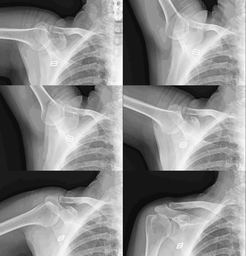
How does Flexion-Extension X-ray work?
Flexion-Extension X-ray is a new form of dynamic digital radiography that was recently FDA approved. Also know as Joint Cinegraphy, Flexion-Extension X-ray allows us to take multiple X-ray images at 6 or 15 frames per second, to be viewed in motion. Unlike alternatives, Cinegrams offer a large, highly detailed 17" x 17" image. There is no extra imaging, radiation or post processing time.
Each frame we acquire is a single diagnostic-quality image, and the entire sequence can be replayed and paused at individual frames for single-frame viewing and diagnostic purposes. DDR support diagnosing musculoskeletal conditions by presenting diagnostic details in full motion to assess the spine and joint stability. With DDR, orthopedists can quantify the dynamic relationship between bones and soft tissue through the full range of motion in weight-bearing or resting positions for faster and more detailed diagnoses.
What are the Benefits of Flexion-Extension X-ray?
- Brand new technology that shows orthopedic range of motion.
- NO extra views needed with all images included in a single exam.
- Less radiation than fluoroscopy exams.
- Extremely beneficial for post-surgical follow up for chiropractors and orthopedists.
- Picks up dynamic instability for lateral cervical spine where other modalities fall short.
During a DDR X-ray, you are typically asked to remove any jewelry to avoid interference with the images. You will then stand, sit, or lie down depending on the body part being scanned. The radiologic technologist will then get the x-ray machine into position and capture clear images. You may be instructed to take a deep breath and hold it briefly while the X-ray is taken to ensure clear images. The entire process is quick and painless, lasting only a few minutes. The images are then reviewed by a radiologist for diagnosis.
Please leave all jewelry at home and wear cloths with no metal if possible.