Ultrasound at ZP
Ultrasound, or sonography, is performed at every ZP location. Our state-of-the-art high resolution ultrasound systems obtain images of internal organs and other soft-tissue structures inside the body. Our certified medical sonographers are dedicated to taking the time necessary to provide quality service to all of our patients. Ultrasound is a safe and painless test that uses high frequency sound waves to produce images. The sound waves cannot be heard or felt. Still and moving real-time images can be captured during an ultrasound.
-

Fastest scans
-

Qualified staff
-

Sharpest images

The Benefits of Ultrasound
- Produces highly detailed images of soft tissues, organs, and structures.
- Safe and radiation-free, making it ideal for patients of all ages, including pregnant individuals
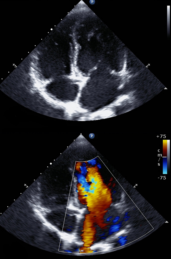
- Real-time imaging that allows providers to observe movement, blood flow, and organ function instantly
- Noninvasive and painless, with no needles or injections required for most exams
- Widely versatile, used to evaluate organs, soft tissues, vessels, and developing pregnancies
- Quick and convenient, typically completed within minutes with no downtime afterward
- Useful at guiding a number of procedures, such as biopsies and arthrograms
- Cost-effective, offering valuable diagnostic information at a lower cost than many other imaging modalities.
-

Most advanced Technology
Fast and noninvasive exams -

Offered at 25 locations
Througout Nassau & Suffolk -

No Radiation
Safe & efficient diagnostics -

Clearest Diagnostic Images
More confidence in your diagnosis
About the Ultrasound Procedure
Ultrasound imaging uses the same principles of SONAR that were developed during World War I for tracking submarines. It was not used for medical purposes until the late 1940s.
To produce images, a transducer, or probe, is placed on your skin and pulses of sound waves are sent through your body. As the sound waves pass through the body, they produce echoes which are received by the transducer and sent to the computer. The echoes are analyzed and converted into images, which in turn create real-time pictures on the monitor. This helps to determine the shape, size, and composition of organs and tissues.
Since ultrasound records images in real time, it is especially useful for examining blood flow and guiding needle biopsy procedures.
Depending on the area being studied, you may be asked to change into a gown.
Before the exam starts, the ultrasound technologist will confirm that any special preparation necessary was followed. You will then be asked to lie down on a comfortably padded examination table.
A small amount of gel will be placed on the area being examined. This gel is harmless and can be easily wiped clean after the exam. The gel prevents any air from getting between the transducer (ultrasound probe) and your skin. This direct contact between the probe and your skin helps the transducer deliver sound waves into your body efficiently.
The ultrasound technologist will place the transducer gently on your skin where the gel was applied and move the probe around slowly. Changing the direction or the angle of the probe allows the sonographer to get the best possible images of the organ or tissue being examined.
Aortic/Abdominal
Nothing to eat, drink, chew, or smoke for six hours prior to your exam.
Breast/Scrotal/Thyroi
No preparation required.
Color Flow Doppler
No preparation required.
Pelvic/Obstetrical
A full bladder is necessary for the exam. Have breakfast and/or lunch. Women must drink at least 32 oz. of water, finishing one hour prior to your exam. Men must drink at least 16 oz. of water, finishing one hour prior to your exam. Do not empty your bladder.
Prostate
Take a fleet enema at least one hour prior to the exam. Have nothing to eat or drink after the fleet enema.
Renal
Drink a 16 oz. glass of water one hour prior to study. Do not void.
Renal Arterial Study
Have nothing to eat, drink, chew, or smoke for six hours prior to your exam. In addition, consult your physician before taking Gas-X one hour before the exam.