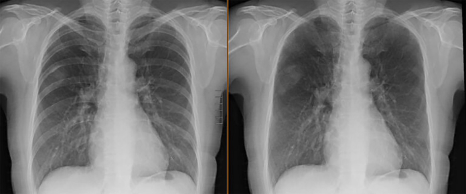
The example above demonstrates a routine chest radiograph (left) paired with the processed bone suppressed image (right).
Bone Suppression Software enhances conventional digital X-ray by creating a second image that digitally minimizes the appearance of the ribs, clavicles, and other bony structures that can obscure the lungs. By providing a clearer view of the lung tissue, this advanced technology helps radiologists detect subtle abnormalities with greater confidence while improving overall diagnostic accuracy. Benefits include:
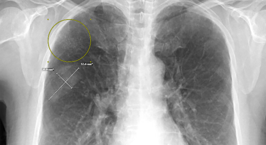
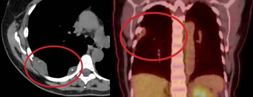
During a chest X-ray with Bone Suppression, you are typically asked to remove any jewelry to avoid interference with the images. You will stand against the X-ray machine, usually facing forward and then sideways. The radiologic technologist will then get the x-ray machine into position and capture clear images of the chest. You will be instructed to take a deep breath and hold it briefly while the X-ray is taken to ensure clear images of the lungs and heart. The entire process is quick and painless, lasting only a few minutes. The images are then reviewed by a radiologist for diagnosis.
Please leave all jewelry at home and wear cloths with no metal if possible.