Body Imaging with MRI at Zwanger-Pesiri
Zwanger-Pesiri Radiology's dedicated Body Imaging team consists of fellowship-trained radiologists and dedicated technologists who specialize in diseases and conditions affecting the major organs and systems in the abdomen, pelvis, and chest. We provide a variety of MRI units to fit your needs, including Wide Bore MRI (3T and 1.5T), Open Sided MRI, and the new metal reduction MRI for patients with metal implanted devices. Read more about our MRI systems.

What is Body MRI?
Body MRI uses state-of-the-art imaging to concentrate on conditions affecting the organs from your neck down to your pelvis. This includes the lungs, liver, stomach, spine, pelvis, kidneys, colon, pancreas, and all portions of the abdomen. MRI is an invaluable tool for body imaging because of its sensitivity to soft tissue, allowing us to visualize organs in great detail. Also, there is no ionizing radiation used during an MRI scan.
Cardiac Magnetic Resonance (CMR) is an advanced form of magnetic resonance imaging that has become very useful for evaluating many cardiovascular conditions. It is used to assess the anatomy, function, and viability of the heart, and can be useful for detecting ischemia and infarction. It can also be helpful for studying valve dysfunction, heart failure, congenital heart disease, masses in and around the heart, and the presence of inherited diseases.
Why might a Body MRI be performed?
We utilize MRI to diagnose and monitor treatments for many conditions in the abdomen, pelvis, and chest, such as tumors. We routinely image the liver for cirrhosis, and digestive tract for ulcerative colitis and Crohn’s disease.
A body MRI can show how well your current treatment is working, and help guide any future treatments. It may also be a useful tool for guiding a biopsy procedure, or for pre-surgical planning.
The body imaging subspecialists at ZP offer expertise for a wide range of exams including:
- General oncologic imaging with MRI.
- Kidney MRI.
- Liver, gallbladder, and biliary system MRI.
- Pancreas MRI.
- Prostate MRI for screening and staging examinations.
- Cardiac MRI.
- Spleen MRI.
- MR angiography for studying blood vessels.
- Breast MRI.
- Urinary bladder MRI.
- Gynecologic cancer MRI for uterus, cervix, and ovaries.
- Rectal cancer staging MRI.
- MR enterography for inflammatory bowel disease.
- Dynamic pelvic/MR defecography MRI.
- Perianal fistula and abscess evaluation MRI.
- Placental and fetal MRI.
Frequently Asked Questions
If you are receiving IV CONTRAST for your exam, have nothing to eat 1 hour prior to your exam time. You may drink clear liquids (example: water, ginger ale, apple juice).
Because the MRI system uses a very powerful magnet, there are many items that cannot be brought into the exam room. Although a private locker will be provided, it is best to leave certain items at home or with someone during your MRI exam, including:
- Cell phones, pagers, iPads, or any other electronic device
- Credit cards, driver’s license, and any other card with a magnetic strip
- Loose change
- Bobby pins, safety pins or any other item with metallic pieces
- Any loose metallic items on clothing or body
- Glasses, hearing aids and items such as TENS Units will not be able to go into the exam room, but can be worn until just before entering the MRI room.
- Wear comfortable loose-fitting clothes, such as a sweatsuit. Be sure there are no metal zippers, snaps or buckles. Do not wear earrings, hairpins, or jewelry. Do not apply eye shadow or mascara.
This exam may not be performed if you have a cardiac pacemaker, cerebral aneurysm clips or a metallic hearing implant. If you are a sheet metal worker or have ever had metal fragments in your eye(s), an orbit x-ray will be taken prior to your MRI exam.
After checking in, you will be escorted to a changing room. You will then meet the registered MRI technologist who will be performing your exam. The technologist will review the screening questionnaire and the procedure with you, answering any questions you may have.
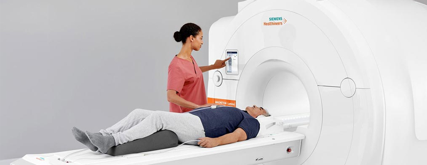
When you are ready, you will be asked to lie down on the cushioned MRI table. Many MRI exams require the use of a coil, which is placed on or around the body part being scanned. The coil is a lightweight device that acts as an antenna to receive the radio frequency signal coming out of your body. That data is then transmitted to a computer, which will then generate the images. If it has been determined that your study requires the use of a contrast material (or dye), the technologist or nurse will perform the injection and administer the iodine-free contrast. Please let the staff know if you have any allergies.
The table will slowly move into position within the large doughnut-shaped magnet. We provide you with noise-canceling headphones and your choice of music, as you will hear a series of loud thumping noises as the scan is performed. With our modern MRI systems, most exams are over in 3 songs! We ask that you keep very still to help ensure that the images come out clear. Throughout the entire exam, you will be able to communicate with the technologist through an intercom. A Cardiac MRI exam can take anywhere from 10 to 30 minutes.