MR Enterography at Zwanger-Pesiri
An MR Enterography is a specialized MRI exam used to obtain detailed images of the gastrointestinal tract. This test is accurate, non-invasive, and is useful for evaluating a broad range of disorders including Crohn's disease. Our fellowship-trained Body Imaging Radiologists are experts in abdominal and pelvic disorders, and work closely with your physician for a quick turnaround and expert interpretation.
Why might I need an MR Enterography exam?
MR Enterography is an effective method for evaluation and diagnosis of disorders of the gastrointestinal tract, including:
- Crohn's disease.
- Polyposis syndromes.
- Inflammatory conditions.
- Celiac disease.
- Diverticular disease.
- Systemic sclerosis.
- Bowel duplication.
MR Enterography may help find:
- Internal bleeding.
- Areas of irritation and swelling.
- Abscesses, which are pus filled pockets, in the intestinal walls.
- Small tears in the intestine wall.
- Blockages.
- MR Enterography may also help track how well certain treatments are working.
Frequently Asked Questions
Have nothing to eat or drink 4 hours prior to exam, except for a sip of water to take normal medications.
Please arrive 1 hour and 15 minutes prior to your scheduled appointment time. You will begin drinking an oral contrast upon arrival.
Refrain from wearing any jewelry or clothes with metal.
After checking in, you will be given an oral contrast to drink. You will then be escorted to a changing room, where you will then meet the registered MRI technologist who will be performing your exam. The technologist will review the screening questionnaire and the procedure with you, answering any questions you may have.
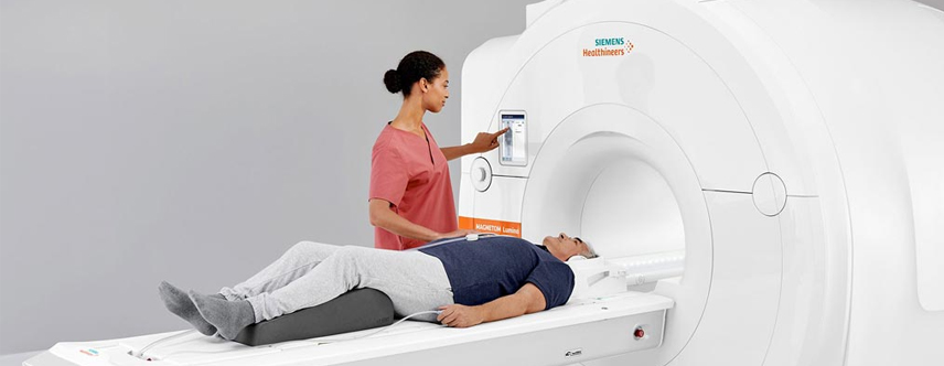
When you are ready, you will be asked to lie down on the cushioned MRI table. Many MRI exams require the use of a coil, which is placed on or around the body part being scanned. The coil is a lightweight device that acts as an antenna to receive the radio frequency signal coming out of your body. That data is then transmitted to a computer, which will then generate the images.
If it has been determined that your study also requires the use of an IV contrast material (or dye), the technologist or nurse will perform the injection and administer the iodine-free contrast. Please let the staff know if you have any allergies.
The table will slowly move into position within the large doughnut-shaped magnet. We provide you with noise-canceling headphones and your choice of music, as you will hear a series of loud thumping noises as the scan is performed. We ask that you keep very still to help ensure that the images come out clear. Throughout the entire exam, you will be able to communicate with the technologist through an intercom. The actual scan can take anywhere from 45 to 60 minutes, depending on the body part being scanned.