Long Island's Brain & Spine Imaging Specialists

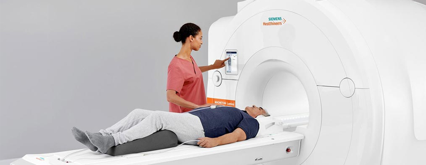
MRI is a noninvasive diagnostic tool that is used to provide extremely detailed images of the brain and spine, as well as to measure blood flow. Our neuroradiological department has decades of experience in neurological MRI, and is here to answer any questions you may have about the procedure. We provide a variety of MRI systems to meet your needs, including the 3T Wide Bore MRI, which is considered the gold standard for imaging the central nervous system. Zwanger-Pesiri invests in the latest MRI technology from Siemens Healthineers.

ZP now offers NeuroQuant®, an AI tool that assists physicians in diagnosing neurogenerative disease of the brain. Click here to learn more about how this software is revolutionizing care for patients with Parkinson's and Alzheimer's disease.

NeuroQuant® for
Neurodegenerative Diseases

NeuroQuant® is advanced AI-powered software that enhances brain MRI examinations by providing precise measurements of brain structures affected by neurodegenerative diseases. Used in the evaluation of conditions such as Alzheimer's disease, Parkinson's disease, and other forms of dementia, NeuroQuant® helps detect subtle changes in brain volume that may be difficult to identify through visual assessment alone. This innovative technology provides physicians with valuable quantitative data, supporting earlier diagnosis, improved disease monitoring, and more informed treatment planning.

What is Neurological MRI used for?

MRI may be used to study the brain or spinal cord for injuries, structural abnormalities, or certain other conditions, such as:

  • Tumor and abscesses in the brain and spinal cord.
  • Eye disease.
  • Inflammation.
  • Infection.
  • Degenerative disorders such as multiple sclerosis.
  • Brain injury from trauma.
  • Subclinical brain edema.
  • Cerebral contusions.
  • Herniations in the spine.
  • Vascular irregularities.
  • Brain damage associated with epilepsy.
  • Weakening and ballooning of an artery (aneurysm).
  • Fluid in the brain (hydrocephalus).
  • Cause of epilepsy (seizures).
  • Pituitary gland disorders.
Neurological MRI

MRI may also be recommended to assist in planning surgeries of the spine, such as a spinal fusion, decompression of a pinched nerve or steroid injection. In addition, it may be used to look for problems after surgery such as infection or scarring.

MR Angiography may be ordered if we are looking specifically at the blood vessels. Our high-tech MRI systems provide superb visualization of the structure and functional changes of the cerebral arteries, and aids in identifying structural anomalies.

MRI exam

Who can benefit from a Cardiac MRI?

A Cardiac MRI may be recommended for patients with:

  • Cardiovascular anatomical anomalies, such as congenital defects.
  • Tumors.
  • Coronary artery disease.
  • Cardiomyopathy.
  • Myocarditis, or covid-induced myocarditis.
  • A history of a heart attack.
  • Cardiac arrhythmia.
  • An enlarged heart, or thickening of the heart muscle.
  • Hemochromatosis.
  • Sarcoid.

Frequently Asked Questions

If you are receiving IV CONTRAST for your exam, have nothing to eat 1 hour prior to your exam time. You may drink clear liquids (example: water, ginger ale, apple juice).

Because the MRI system uses a very powerful magnet, there are many items that cannot be brought into the exam room. Although a private locker will be provided, it is best to leave certain items at home or with someone during your MRI exam, including:

  • Cell phones, pagers, iPads, or any other electronic device
  • Credit cards, driver’s license, and any other card with a magnetic strip
  • Loose change
  • Bobby pins, safety pins or any other item with metallic pieces
  • Any loose metallic items on clothing or body
  • Glasses, hearing aids and items such as TENS Units will not be able to go into the exam room, but can be worn until just before entering the MRI room.
  • Wear comfortable loose-fitting clothes, such as a sweatsuit. Be sure there are no metal zippers, snaps or buckles. Do not wear earrings, hairpins, or jewelry. Do not apply eye shadow or mascara.

This exam may not be performed if you have a cardiac pacemaker, cerebral aneurysm clips or a metallic hearing implant. If you are a sheet metal worker or have ever had metal fragments in your eye(s), an orbit x-ray will be taken prior to your MRI exam.

After checking in, you will be escorted to a changing room. You will then meet the registered MRI technologist who will be performing your exam. The technologist will review the screening questionnaire and the procedure with you, answering any questions you may have.

When you are ready, you will be asked to lie down on the cushioned MRI table. Many MRI exams require the use of a coil, which is placed on or around the body part being scanned. The coil is a lightweight device that acts as an antenna to receive the radio frequency signal coming out of your body. That data is then transmitted to a computer, which will then generate the images. If it has been determined that your study requires the use of a contrast material (or dye), the technologist or nurse will perform the injection and administer the iodine-free contrast. Please let the staff know if you have any allergies.

The table will slowly move into position within the large doughnut-shaped magnet. We provide you with noise-canceling headphones and your choice of music, as you will hear a series of loud thumping noises as the scan is performed. With our modern MRI systems, most exams are over in 3 songs! We ask that you keep very still to help ensure that the images come out clear. Throughout the entire exam, you will be able to communicate with the technologist through an intercom.

An MR Angiography exam can take anywhere from 10 to 30 minutes.