Orthopedic MRI at Zwanger-Pesiri
Long Island's Leader in Orthopedic MRI
When you're dealing with joint pain, a sports injury, back pain, or limited mobility, getting the right diagnosis quickly is the first step toward feeling better. At Zwanger-Pesiri Radiology, we have been a trusted leader in orthopedic MRI on Long Island for decades, providing advanced imaging that helps physicians accurately diagnose musculoskeletal injuries and conditions.
Our fellowship-trained musculoskeletal (MSK) radiologists specialize exclusively in imaging of the bones, joints, muscles, tendons, ligaments, cartilage, and spine. They work closely with orthopedic surgeons, sports medicine physicians, pain management specialists, rheumatologists, podiatrists, and primary care providers to deliver accurate, detailed interpretations that guide the best treatment plan.
We combine this expertise with state-of-the-art MRI technology, including 3T Wide Bore MRI, the gold standard for orthopedic imaging. Our advanced MRI systems produce exceptional image quality with greater detail, faster scan times, and a more comfortable experience for patients.
Whether you're recovering from an injury, living with chronic joint pain, or preparing for orthopedic surgery, Zwanger-Pesiri provides the high-quality imaging your physician needs to help you move forward with confidence.
What is Orthopedic MRI?
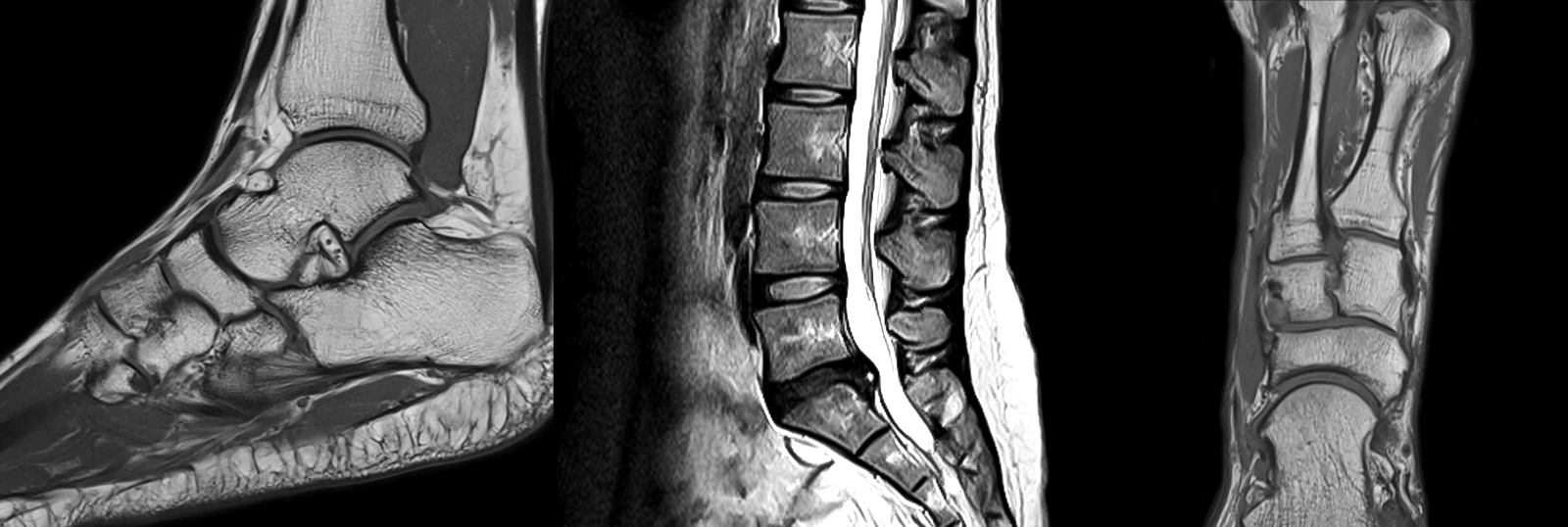
An orthopedic MRI, also known as a musculoskeletal (MSK) MRI, is a noninvasive imaging exam that creates highly detailed images of the body's bones, joints, cartilage, muscles, tendons, ligaments, and other soft tissues. Unlike X-rays or CT scans, MRI does not use ionizing radiation.
MRI is one of the most accurate ways to diagnose orthopedic injuries and conditions because it allows physicians to see structures that cannot be evaluated on standard X-rays.
An orthopedic MRI may be recommended to:
- Identify the cause of joint or muscle pain
- Diagnose sports injuries
- Evaluate ligament, tendon, cartilage, and meniscus tears
- Detect stress fractures and hidden bone injuries
- Assess arthritis and joint degeneration
- Evaluate neck and back pain
- Plan orthopedic surgery
- Monitor healing after surgery or injury
Early and accurate diagnosis often leads to faster treatment and better outcomes.

MRI for Sports Injuries
Whether you're a professional athlete, weekend warrior, or simply enjoy staying active, sports injuries require an accurate diagnosis before treatment begins.
MRI is the preferred imaging exam for many athletic injuries because it provides exceptional detail of the muscles, ligaments, tendons, cartilage, and joints that are commonly injured during sports and physical activity.
Our fellowship-trained musculoskeletal radiologists specialize in sports medicine imaging and routinely diagnose:
- ACL and MCL tears
- Meniscus injuries
- Rotator cuff tears
- Labral tears
- Achilles tendon injuries
- Muscle strains
- Stress fractures
- Cartilage damage
- Overuse injuries
We work closely with your orthopedic surgeon, sports medicine physician, or referring provider to determine the severity of your injury, guide treatment decisions, and help support a safe return to activity while reducing the risk of reinjury.
Frequently Asked Questions
If you are receiving IV CONTRAST for your exam, have nothing to eat 1 hour prior to your exam time. You may drink clear liquids (example: water, ginger ale, apple juice).
Because the MRI system uses a very powerful magnet, there are many items that cannot be brought into the exam room. Although a private locker will be provided, it is best to leave certain items at home or with someone during your MRI exam, including:
- Cell phones, pagers, iPads, or any other electronic device
- Credit cards, driver’s license, and any other card with a magnetic strip
- Loose change
- Bobby pins, safety pins or any other item with metallic pieces
- Any loose metallic items on clothing or body
- Glasses, hearing aids and items such as TENS Units will not be able to go into the exam room, but can be worn until just before entering the MRI room.
- Wear comfortable loose-fitting clothes, such as a sweatsuit. Be sure there are no metal zippers, snaps or buckles. Do not wear earrings, hairpins, or jewelry. Do not apply eye shadow or mascara.
This exam may not be performed if you have a cardiac pacemaker, cerebral aneurysm clips or a metallic hearing implant. If you are a sheet metal worker or have ever had metal fragments in your eye(s), an orbit x-ray will be taken prior to your MRI exam.
After checking in, you will be escorted to a changing room. You will then meet the registered MRI technologist who will be performing your exam. The technologist will review the screening questionnaire and the procedure with you, answering any questions you may have.
When you are ready, you will be asked to lie down on the cushioned MRI table. Many MRI exams require the use of a coil, which is placed on or around the body part being scanned. The coil is a lightweight device that acts as an antenna to receive the radio frequency signal coming out of your body. That data is then transmitted to a computer, which will then generate the images. If it has been determined that your study requires the use of a contrast material (or dye), the technologist or nurse will perform the injection and administer the iodine-free contrast. Please let the staff know if you have any allergies.
The table will slowly move into position within the large doughnut-shaped magnet. We provide you with noise-canceling headphones and your choice of music, as you will hear a series of loud thumping noises as the scan is performed. With our modern MRI systems, most exams are over in 3 songs! We ask that you keep very still to help ensure that the images come out clear. Throughout the entire exam, you will be able to communicate with the technologist through an intercom.
An Orthopedic MRI exam can take anywhere from 10 to 30 minutes.