Cardiac MRI (Heart MRI) at Zwanger-Pesiri
Advanced Cardiac MRI on Long Island
When your physician needs the most detailed information about your heart, a Cardiac MRI, also known as a Heart MRI, is one of the most advanced imaging tests available. This noninvasive exam provides highly detailed images of your heart's structure, function, blood flow, and heart muscle, helping physicians diagnose a wide range of cardiovascular conditions with exceptional accuracy.
At Zwanger-Pesiri Radiology, we are proud to be a leader in advanced Cardiac MRI on Long Island. Our experienced cardiac imaging team works closely with your cardiologist to ensure a seamless experience—from scheduling your exam to delivering timely, accurate results. Whether you are being evaluated for chest pain, heart failure, cardiomyopathy, myocarditis, or another heart condition, our team is committed to providing the answers you and your physician need.
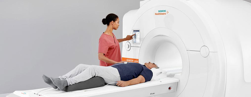
Using the latest 3T and 1.5T Wide Bore MRI technology from Siemens Healthineers, we deliver exceptional image quality, shorter scan times, and a more comfortable patient experience.

What Is a Cardiac MRI (Heart MRI)?
A Cardiac MRI, sometimes called a Heart MRI, is a noninvasive imaging exam that uses a powerful magnetic field and radio waves to create highly detailed pictures of your heart. Unlike CT scans and X-rays, Cardiac MRI does not use ionizing radiation.
A Cardiac MRI allows physicians to evaluate:
- Heart muscle health
- Blood flow through the heart
- Heart chamber size
- Heart valve function
- Damage after a heart attack
- Inflammation of the heart muscle
- Scar tissue (fibrosis)
- Congenital heart defects
- Cardiac tumors and masses
- Your heart's pumping function (ejection fraction)
Because it provides information that other imaging tests often cannot, Cardiac MRI is considered one of the most comprehensive and accurate ways to evaluate many heart conditions.
Why Might You Need a Cardiac MRI?
Your physician may recommend a Cardiac MRI to diagnose or monitor conditions such as:
- Cardiomyopathy
- Myocarditis, including inflammation following viral illnesses or COVID-19
- Coronary artery disease
- Heart failure
- Previous heart attack
- Cardiac arrhythmias
- Enlarged heart
- Thickened heart muscle (hypertrophic cardiomyopathy)
- Heart valve disease
- Congenital heart defects
- Cardiac tumors or masses
- Sarcoidosis affecting the heart
- Hemochromatosis (iron overload)
Cardiac MRI is also commonly recommended when your physician needs additional information following an echocardiogram, CT scan, or other cardiac imaging study.
Why Choose Cardiac MRI?
Exceptional Accuracy
Cardiac MRI is considered the gold standard for measuring ejection fraction, one of the most important indicators of how effectively your heart pumps blood.
Outstanding Heart Muscle Imaging
Cardiac MRI provides exceptional visualization of the heart muscle, allowing physicians to detect inflammation, scar tissue, fibrosis, tumors, and areas of damaged or healthy heart muscle with remarkable precision.
No Radiation Exposure
Unlike CT scans and many nuclear medicine studies, Cardiac MRI uses no ionizing radiation, making it an excellent choice for many patients.
Advanced Functional Imaging
Using specialized cardiac gating and advanced CINE MRI technology, Cardiac MRI captures moving images of your beating heart. This allows physicians to evaluate:
Heart muscle contraction- Blood flow through the heart
- Valve movement
- Overall heart function
- Ejection fraction
These detailed images provide critical information for diagnosis and treatment planning.
What to Expect During Your Cardiac MRI
During your examination, you will lie comfortably on the MRI table while detailed images of your heart are obtained. Small electrodes placed on your chest synchronize image acquisition with your heartbeat, allowing us to capture clear, precise images throughout the cardiac cycle.
Depending on your examination, intravenous contrast may be used to evaluate heart muscle viability, scar tissue, inflammation, or blood flow.
Most Cardiac MRI examinations take approximately 45 to 60 minutes.
Our experienced MRI technologists will guide you through every step of the process and work to ensure you remain comfortable throughout your exam.
Why Choose Zwanger-Pesiri?
Patients and physicians throughout Long Island trust Zwanger-Pesiri for advanced cardiac imaging because we offer:
- Specialized Cardiac MRI expertise
- Close collaboration with your cardiologist
- Advanced Siemens Healthineers MRI technology
- 3T and 1.5T Wide Bore MRI systems
- Outstanding image quality with shorter scan times
- Comfortable outpatient imaging centers
- Convenient Long Island locations
- Comprehensive evaluation of heart structure, heart muscle health, blood flow, and cardiac function
Depending on your examination, intravenous contrast may be used to evaluate heart muscle viability, scar tissue, inflammation, or blood flow.
At Zwanger-Pesiri Radiology, our mission is to provide the highest-quality Cardiac MRI in a comfortable outpatient setting, delivering the detailed information your physician needs to diagnose heart disease early, personalize treatment, and help you achieve the best possible heart health.
Frequently Asked Questions
If you are receiving IV CONTRAST for your exam, have nothing to eat 1 hour prior to your exam time. You may drink clear liquids (example: water, ginger ale, apple juice).
Because the MRI system uses a very powerful magnet, there are many items that cannot be brought into the exam room. Although a private locker will be provided, it is best to leave certain items at home or with someone during your MRI exam, including:
- Cell phones, pagers, iPads, or any other electronic device
- Credit cards, driver’s license, and any other card with a magnetic strip
- Loose change
- Bobby pins, safety pins or any other item with metallic pieces
- Any loose metallic items on clothing or body
- Glasses, hearing aids and items such as TENS Units will not be able to go into the exam room, but can be worn until just before entering the MRI room.
- Wear comfortable loose-fitting clothes, such as a sweatsuit. Be sure there are no metal zippers, snaps or buckles. Do not wear earrings, hairpins, or jewelry. Do not apply eye shadow or mascara.
This exam may not be performed if you have a cardiac pacemaker, cerebral aneurysm clips or a metallic hearing implant. If you are a sheet metal worker or have ever had metal fragments in your eye(s), an orbit x-ray will be taken prior to your MRI exam.