Magnetic Resonance Enterography (MRE) is a non-invasive diagnostic tool used to visualize the small intestine and assess its function. It is a safe and effective alternative to traditional diagnostic methods such as barium X-rays or endoscopy. At Zwanger-Pesiri, our expert radiologists use state-of-the-art MRI equipment to perform MR Enterography, delivering accurate results and a high level of patient comfort.
What Is MR Enterography?
MR Enterography is a type of Magnetic Resonance Imaging (MRI) scan that specifically focuses on the small intestine. Unlike traditional X-rays, MRI uses magnetic fields and radio waves to produce detailed images of the body's internal structures. During an MR Enterography exam, the patient is positioned on the MRI table and a special contrast material is introduced into the small intestine to enhance the visibility of the intestinal walls and surrounding tissues.
MR Enterography is typically ordered by a physician to evaluate symptoms such as abdominal pain, diarrhea, weight loss, or to assess the status of pre-existing conditions such as Crohn's disease or small intestine tumors. This exam is particularly useful for detecting changes in the small intestine that may be indicative of these conditions.


Your Comfort During The Process
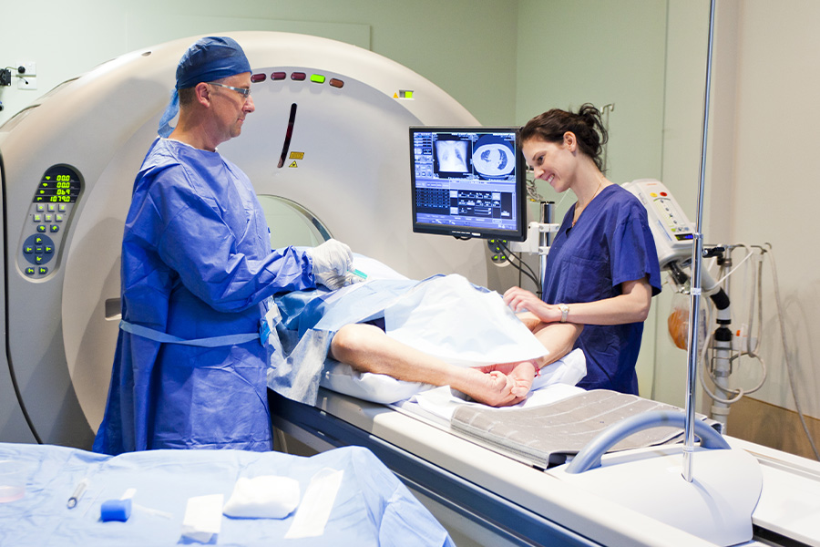
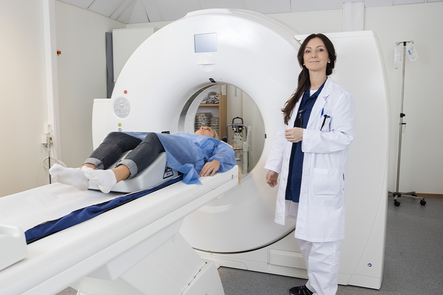
At Zwanger-Pesiri, our team of experts provides a comfortable and efficient MR Enterography experience. Before the exam, the patient will be asked to remove any metal objects such as jewelry, clothing with metal zippers, or hearing aids. They will then be positioned on the MRI table and a special contrast material will be introduced into the small intestine through a tube or enema. The MRI machine will then be used to produce detailed images of the small intestine, which our radiologists will review to determine the presence of any abnormal conditions.
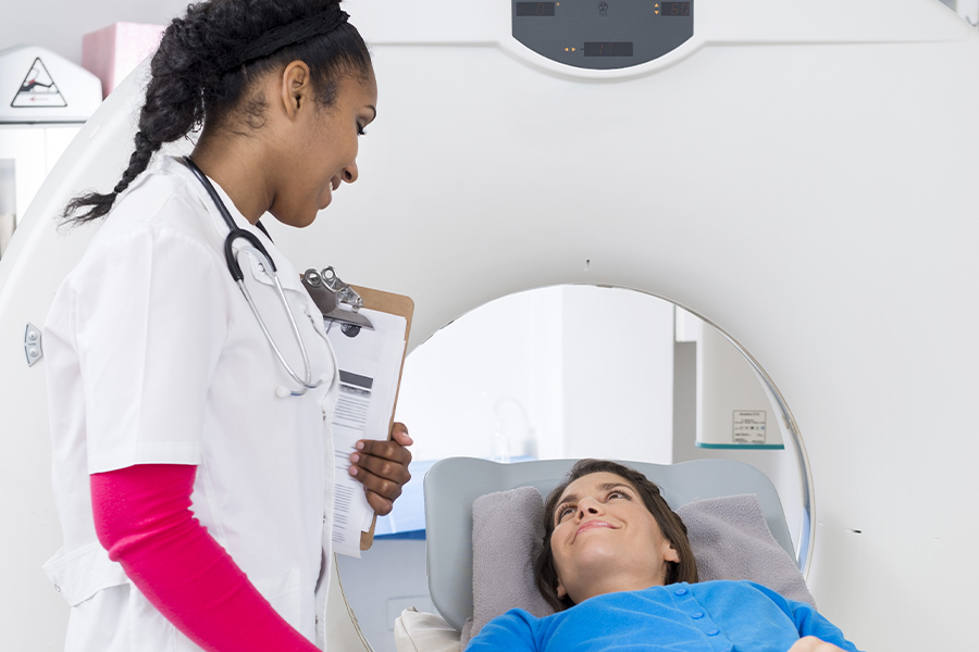
The MR Enterography exam usually takes about 30-60 minutes, during which time the patient will lie still on the MRI table. The examiner will be in constant communication with the patient during the exam, making sure they are comfortable and aware of what is happening. After the exam, the patient will be able to return to their normal activities immediately, with no downtime or recovery time required. The results of the MR Enterography exam will be reviewed by one of our experienced radiologists and a report will be provided to the referring physician for further evaluation and treatment planning if necessary.
It’s A Safe Procedure
MR Enterography is a safe and non-invasive diagnostic tool. Unlike X-rays, it does not use ionizing radiation, making it a safer option for patients who may be pregnant or have pre-existing medical conditions. The contrast material used during the exam is also considered safe, with few reported side effects.
MR Enterography is a highly accurate diagnostic tool that provides detailed images of the small intestine, allowing for a comprehensive evaluation of its structure and function. This exam can be especially helpful for patients who have difficulty undergoing more invasive diagnostic procedures, as it is a non-invasive alternative that provides valuable information with minimal discomfort.

For those looking to undergo MR Enterography, we invite you to schedule an appointment with us at Zwanger-Pesiri. Our team of experienced professionals is here to answer any questions and provide you with the highest level of care. We look forward to helping you achieve optimal health and wellness, so contact us today!
