Ultrasound
Ultrasound at ZP
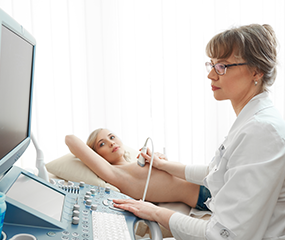
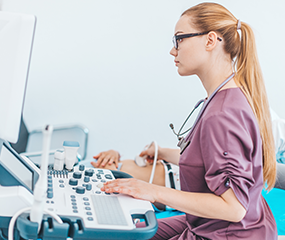
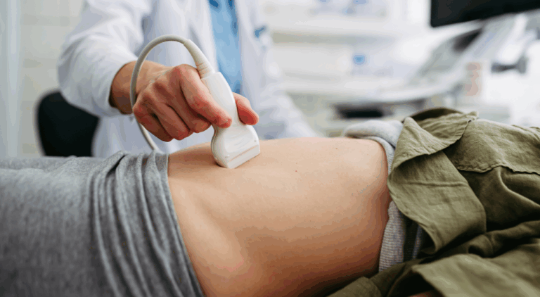
Ultrasound, or sonography, is performed at every ZP location. Our state-of-the-art high resolution ultrasound systems obtain images of internal organs and other soft-tissue structures inside the body. Our certified medical sonographers are dedicated to taking the time necessary to provide quality service to all of our patients. Ultrasound is a safe and painless test that uses high frequency sound waves to produce images. The sound waves cannot be heard or felt. Still and moving real-time images can be captured during an ultrasound.

What are the benefits of Ultrasound?
- Safe and radiation-free, making it ideal for patients of all ages, including pregnant individuals.
- Real-time imaging that allows providers to observe movement, blood flow, and organ function instantly.
- Noninvasive and painless, with no needles or injections required for most exams.
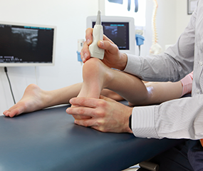
- Widely versatile, used to evaluate organs, soft tissues, vessels, and developing pregnancies.
- Quick and convenient, typically completed within minutes with no downtime afterward.
- Useful at guiding a number of procedures, such as biopsies and arthrograms.
- Cost-effective, offering valuable diagnostic information at a lower cost than many other imaging modalities.

Learn About the Different Types of Ultrasound Exams
 Breast ultrasound (or breast sonogram) is one of the safest and most decisive imaging options available today. They are useful for evaluating nodules, lumps, dense breasts, and more and can differentiate between fluid-filled cysts and solid masses. Commonly they are used along with mammograms to screen for breast cancer or as a guide for biopsies.
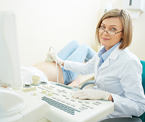

Noninvasive, safe, and does not use any radiation

Widely available and less expensive than other imaging methods

Clear pictures of soft tissues that don’t show up well on x-rays

Real-time imaging for guiding minimally-invasive procedures

Elastografía Hepática
Ahora ofrecemos exámenes de ultrasonido con Elastografía Hepática El avanzado departamento de ultrasonido de…

Mamografía 3D
Mamografía 3D en ZP Su mamografía de detección anual es uno de los pasos más…

Food Donation to Pronto of Long Island
Zwanger-Pesiri Radiology, Long Island’s leading provider of diagnostic imaging services, is proud to announce the…
ZP Hauppauge Grand Opening
Zwanger-Pesiri Radiology is proud to announce the grand opening of our newest state-of-the-art location in…

Best of Long Island 2025
Zwanger-Pesiri Radiology is deeply honored and grateful to the Long Island community for voting us…
Trophon® at ZP
Every ZP location uses Trophon® on ultrasound probes. Trophon® is a breakthrough disinfection technology for ultrasound probe reprocessing. With the ever increasing challenges in the fight against the spread of Healthcare Acquired Infections (HAIs), Trophon’s powerful disinfection technology is setting a new benchmark in protecting patients. At the same time it safeguards staff and the environment from the dangerous hazards and toxic side effects of traditional disinfection methods.
Why Choose Zwanger Pesiri?
Zwanger-Pesiri Radiology brings world-class expertise to the Long Island community. Our subspecialty-trained radiologists are Board Certified by the American Board of Radiology with fellowship training in a variety of specialties. They are highly-skilled, highly-knowledgeable, and make patient care a priority. To learn more, contact us today.