Dynamic Pelvic MRI/MR Defecography
When the pelvic floor muscles and ligaments supporting a woman’s pelvic organs weaken, the
pelvic organs can drop out of place. This is a common condition called pelvic organ prolapse.
Most women develop prolapse after menopause, childbirth, or hysterectomy. It can be
debilitating as well as embarrassing and can significantly impact the quality of life. The most
common symptoms of pelvic organ prolapse are urinary incontinence, incomplete bladder
emptying, pelvic pain, constipation, obstructed and difficult defecation, and fecal incontinence.

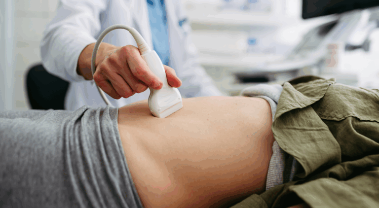
What is Dynamic Pelvic Floor MRI and MR Defecography?
Until recently, fluoroscopic defecography was used for pelvic floor imaging, which exposed patients to radiation (x-rays). With rapid advances in MRI technology, both functional and anatomic evaluations can be performed in a single examination without using radiation. MRI allows us to image in any plane, free from the confusion of overlying structures, and can detect structural abnormalities such as tumors, abscesses, hernias, and defects or lesions in the pelvic floor muscle. These findings can help you and your doctor plan for the best clinical treatment or surgery.
What are the benefits of Dynamic Pelvic Floor MRI and MR Defecography?
-
Provides real-time visualization of pelvic floor muscles and organ movement during various functional maneuvers.
-
Accurately assesses pelvic floor disorders including prolapse, rectocele, enterocele, anismus, and incontinence.
-
Offers superior soft-tissue detail compared to traditional fluoroscopic defecography.
-
Helps pinpoint the exact cause of pelvic pain, pressure, or bowel dysfunction for more targeted treatment.
-
Completely noninvasive and radiation-free, making it a safe diagnostic option.
-
Allows evaluation of multiple pelvic compartments, giving a comprehensive view of overall pelvic floor function.
-
Guides treatment planning for urogynecology, colorectal surgery, and pelvic floor physical therapy.

What Happens During the Procedure?
- You will be asked to perform Kegel exercises while images are taken. This is when you tighten the pelvic floor muscles, similar to stopping the urine flow while urinating.
- You will be asked to perform the Valsalva maneuver by taking a deep breath and bearing down without letting the gel out while images are taken. You may be asked to repeat this step.
- You will be asked to perform defecation while images are taken. You will take a deep breath and bear down towards the rectum and push the contrast gel out of the rectum. You may be asked to repeat this step.
Elastografía Hepática
Ahora ofrecemos exámenes de ultrasonido con Elastografía Hepática El avanzado departamento de ultrasonido de…

Mamografía 3D
Mamografía 3D en ZP Su mamografía de detección anual es uno de los pasos más…

Food Donation to Pronto of Long Island
Zwanger-Pesiri Radiology, Long Island’s leading provider of diagnostic imaging services, is proud to announce the…
ZP Hauppauge Grand Opening
Zwanger-Pesiri Radiology is proud to announce the grand opening of our newest state-of-the-art location in…

Best of Long Island 2025
Zwanger-Pesiri Radiology is deeply honored and grateful to the Long Island community for voting us…
Why Choose Zwanger-Pesiri?
Zwanger-Pesiri Radiology brings world-class expertise to the Long Island community. Our subspecialty-trained radiologists are Board Certified by the American Board of Radiology with fellowship training in a variety of specialties. They are highly-skilled, highly-knowledgeable, and make patient care a priority. To learn more, contact us today.