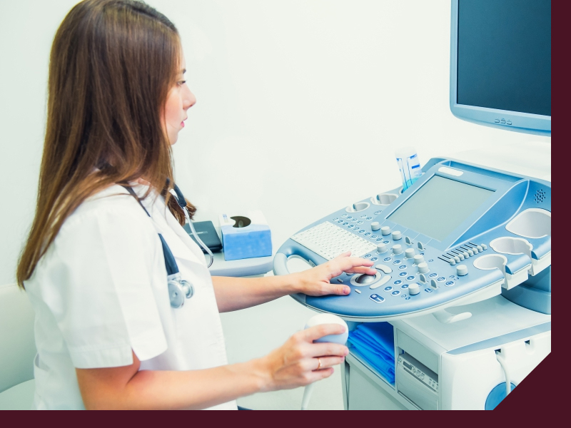
Our expert radiologists work diligently to give you clear, concise, and accurate radiology scans for your MRI, CT, PET, X-ray, ultrasound, biopsy, and more. All of our diagnostic imaging procedures involve a highly-trained radiologist of Zwanger-Pesiri to view the image and report the finding to the patient's referring physician who will then discuss these results during the follow-up visit. Our mission is to help improve the health of our community, setting the gold standard in patient care and technology. Our subspecialized radiologists are highly-trained, working with staff to ensure an unmatched level of satisfaction.
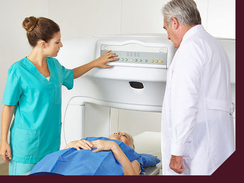
MRI
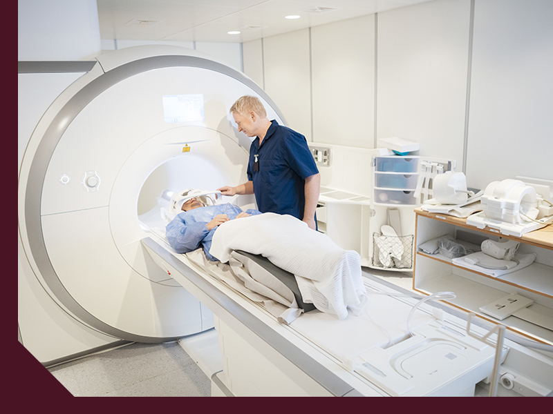
Using a strong magnetic field and radio waves, an MRI produces detailed images of organs and tissues. The atoms in your body respond to this energy, and the MRI detects this response. Unlike X-rays or CT exams, an MRI does not use radiation.
CT
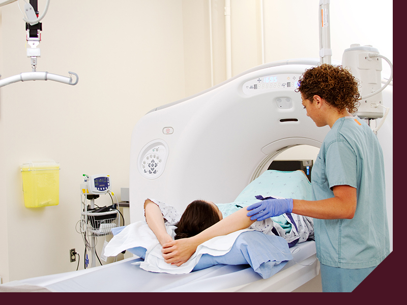
CT is used to help diagnose a range of conditions, including infections, cancer, heart disease, internal injury, musculoskeletal disorders, or gastrointestinal disease. CT scans only use a small amount of X-ray radiation to generate images, but each patient’s exam is individualized to ensure the best possible image while minimizing the amount of radiation.
Calcium Scoring
Cardiac calcium scoring detects a build-up of plaque, which is made of fat, calcium, and other substances, that can narrow or close the arteries. This is a non-invasive CT scan of the heart that calculates your risk of developing coronary artery disease, or CAD, by measuring the amount of calcified plaque in the coronary arteries. A calcium score is calculated based on the amount of plaque observed in the CT scan. It can be converted to a percentile rank based on your age and gender.
Ultrasound
Ultrasound imaging, also called ultrasound scanning or sonography, is a method of obtaining images from inside the body through the use of high-frequency sound waves. No radiation is involved in ultrasound imaging. The reflections of the wave echoes are recorded and displayed as real-time visual images. Ultrasound is a useful way to examine many of the body’s internal organs, including the liver, gallbladder, kidneys, uterus, ovaries, aorta, and other major blood vessels.

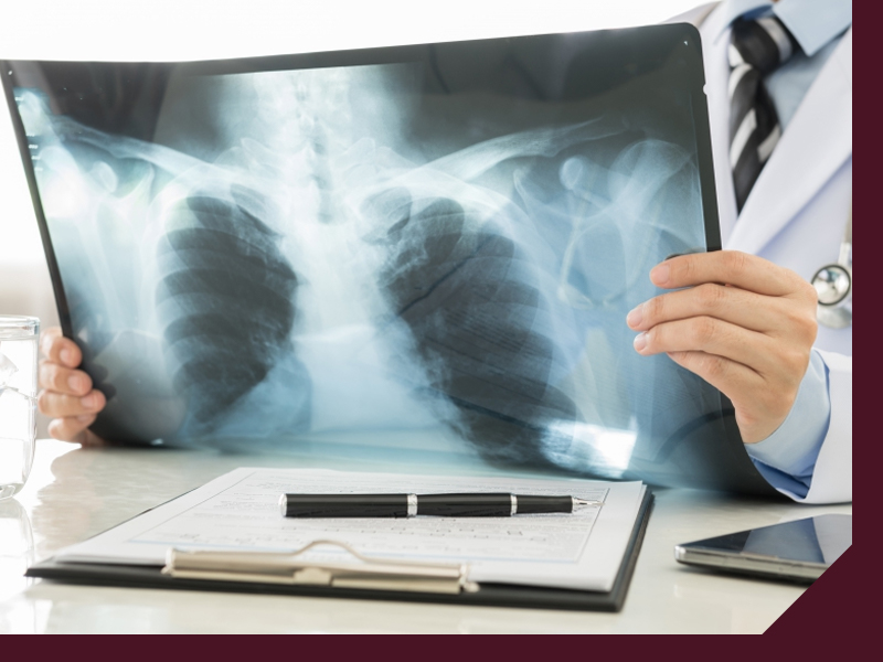
X-Ray
X-rays use invisible electromagnetic energy beams to produce images of internal tissues, bones, and organs on film or digital media. X-rays use external radiation to produce images of the body and can be performed for many reasons, including diagnosing tumors or bone injuries.
Biopsy
A patient may undergo a biopsy when cells or tissue need to be closely examined under a microscope to see if any kind of disease or abnormality is present. Some biopsies only involve removing a small amount of tissue sample with a need while others involve surgically removing a suspicious lump or nodule. Most biopsies are performed on an outpatient basis with minimal preparation, but your doctor will give your clear and direct instruction based on the type of the biopsy being performed.
