X-Ray
X-Ray at ZP
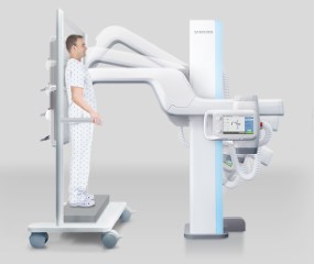
Zwanger-Pesiri offers digital X-ray technology at all of our locations. Our extremely flexible units offer the most comfortable exams with the least amount of radiation. Virtually all of the views can be done upright, which is more comfortable for patients. Our X-ray tables can hold up to 495 lbs.
ZP participates in Image Wisely™, a campaign that encourages smart medical imaging. We pledge to eliminate unnecessary scans and to lower radiation doses by using state-of-the-art equipment for every study at every location. X-rays can either be scheduled or performed on a walk-in basis.
Notify the technologist if you are pregnant or think you may be pregnant. Other than that, there are no preparations for X-ray exams.

What are the Benefits of the latest X-ray technology?
-
Quick and efficient imaging, allowing for rapid diagnosis and treatment decisions
-
Low radiation exposure, especially with modern digital X-ray systems
-
High diagnostic value for evaluating bones, lungs, and certain soft tissues
-
Noninvasive and painless, making exams comfortable for patients
-
Widely available and cost-effective compared to many advanced imaging modalities
-
Ideal for detecting fractures, infections, arthritis, and lung conditions

How does X-ray work?
During an X-ray, beams are produced by a special X-ray tube which is like a supercharged light bulb. The beams are sent through the body to highly specialized digital sensor plates. These plates are similar to those in a digital camera but are much larger. Different parts of the body absorb the X-rays in varying degrees. Dense materials like bones appear white on an X-ray, while soft tissue such as fat and muscle are varying shades of gray. Lungs are mostly filled with air and appear almost black.
A computer then processes and records the images taken without any film being utilized. These electronically stored images are easily accessible and can be compared to other X-ray studies as needed.

What are Some Types of X-ray Exams?
 Chest X-ray uses a very small dose of ionizing radiation to produce pictures of the inside of the chest. It is used to evaluate the lungs, heart, and chest wall and may be used to help diagnose shortness of breath, persistent cough, fever, chest pain, or injury. It also may be used to help diagnose and monitor treatment for a variety of lung conditions such as pneumonia, emphysema, and cancer. Because chest X-ray is fast and easy, it is particularly useful in emergency diagnosis and treatment.
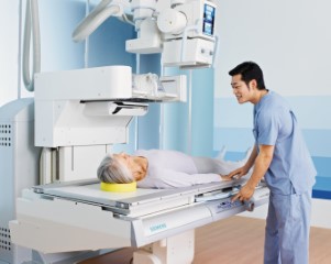 Fluoroscopy is a study of moving body structures — similar to an X-ray "movie." A continuous X-ray beam is passed through the body part being examined. The beam is transmitted to a TV-like monitor so that the body part and its motion can be seen in detail. Fluoroscopy, as an imaging tool, enables physicians to look at many body systems, including the skeletal, digestive, urinary, respiratory, and reproductive systems.
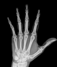 X-ray has become an integral part of imaging bones throughout the body. X-ray uses a very small dose of ionizing radiation, and is commonly used to diagnose fractured bones or joint dislocation. Bone X-rays are the fastest and easiest way for your doctor to view and assess bone fractures, injuries, and joint abnormalities.
During the Test
Our technologist will position your body based on the type of X-ray being performed. Depending on the exam, you will be asked to stand, sit, or lie down on the X-ray table. A lead apron may be placed over your pelvis or breasts to protect you from radiation. The part of your body being evaluated will be exposed to radiation for a fraction of a second to create the image. Our radiologic technologists are trained to use the least amount of radiation possible to produce an accurate image that will help with diagnosis. In addition, our modern X-ray systems have tightly controlled X-ray beams and dose control methods to minimize radiation. This keeps patient exposure to radiation at a minimum.
Diagnostic Radiologists
Board Certified, Fellowship Trained

Why Radiologist Choice Matters
Radiology is an important department in the medical field. From x-rays and ultrasounds to diagnostic…

4 Reasons Why Diagnostic Imaging Is Important
When it comes to the medical industry, there are a lot of moving parts. You…

Women’s Top 4 Health Concerns
Taking care of your health is vital if you want to live a long, full…

Healthy Mind, Healthy Body
When many of us think about health, we are concerned about our physical health needs.…

June: Men’s Health Awareness Month
June is known as men’s health awareness month and at Zwanger Pesiri in Long Island,…
Why Choose Zwanger-Pesiri?
Zwanger-Pesiri Radiology brings world-class expertise to the Long Island community. Our subspecialty-trained radiologists are Board Certified by the American Board of Radiology with fellowship training in a variety of specialties. They are highly-skilled, highly-knowledgeable, and make patient care a priority. To learn more, contact us today.