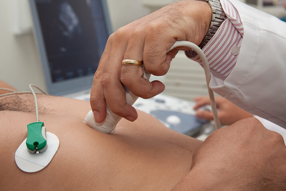
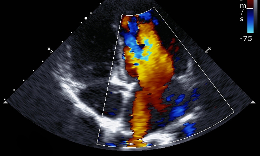
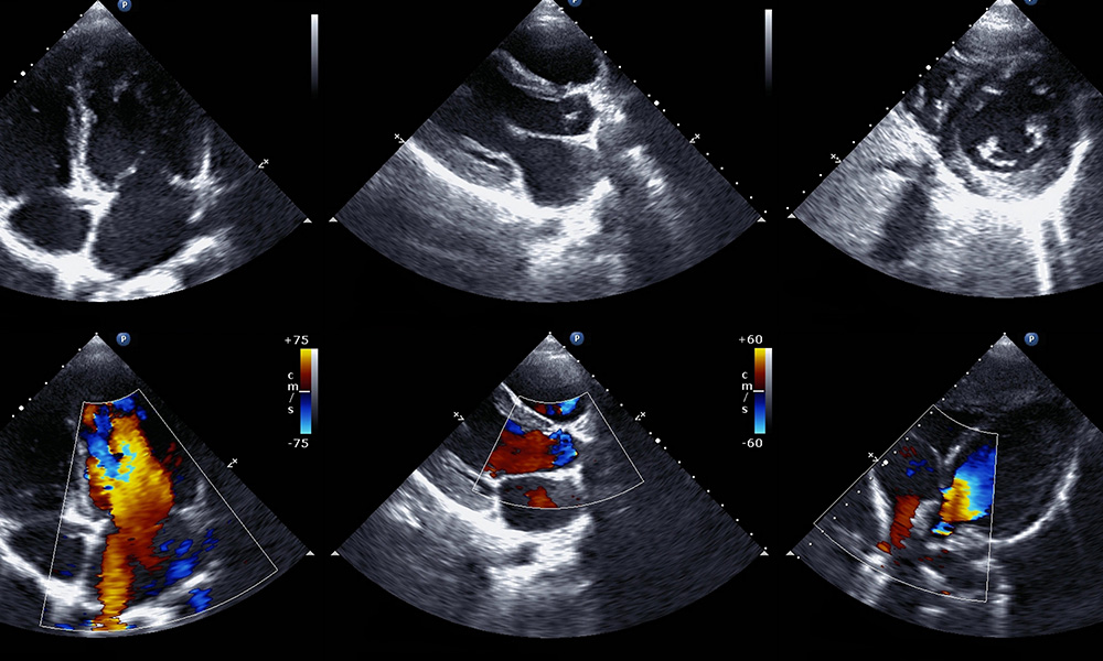
An echocardiogram is an ultrasound test that uses sound waves to produce images of your heart. This test allows us to see your heart beating and blood being pumped. Your doctor can use the images from an echocardiogram to identify heart disease.
Our dedicated cardiac ultrasound department has extensive experience in echocardiography and is here to speak directly with your physician for the best patient outcomes.

What is an Echocardiogram?
An echocardiogram uses ultrasound technology, along with electrodes, to check your heart’s rhythm and to see how blood moves through your heart. An echocardiogram can help your doctor diagnose certain types of heart conditions. It allows us to figure out if your heart's chambers and valves are pumping blood through your heart. The sound waves from the ultrasound make moving pictures of your heart to get a good look at its size and shape.

Why might I need an echocardiogram?
Your doctor may order an echocardiogram to:
- Look for heart disease
- Monitor heart valve disease over time
- See how well medical or surgical treatments are working
- Check for problems with the valves or chambers of your heart
- Check if heart problems are the cause of symptoms such as shortness of breath or chest pain
- Detect congenital heart defects before birth (fetal echocardiogram)


A TTE exam for the heart is the most common type of echocardiogram performed for those with heart conditions. Transthoracic means on the chest wall. An echocardiogram, or echo, is an ultrasound test of the heart. During a transthoracic echo, we use sound waves to create computerized outlines of your heart and its attached blood vessels. This allows us to visualize your heart’s chambers, valves and blood vessels for cardiac issues including extra fluid building up around your heart.
